Is that new dark spot on your skin a harmless age spot or something that requires immediate medical attention? While melasma and age spots represent benign pigmentation, melanoma can initially resemble harmless discoloration. A good dermatologist in Singapore can differentiate between cosmetic concerns and medical conditions requiring attention.
Pigmentation changes occur through melanin production alterations, sun exposure effects, hormonal fluctuations, or cellular mutations. Normal pigmentation typically develops gradually and maintains consistent characteristics. Concerning lesions often display asymmetry, irregular borders, multiple colors, or rapid changes. Understanding these distinctions helps identify when professional evaluation becomes necessary.
Normal Pigmentation Patterns
Melasma
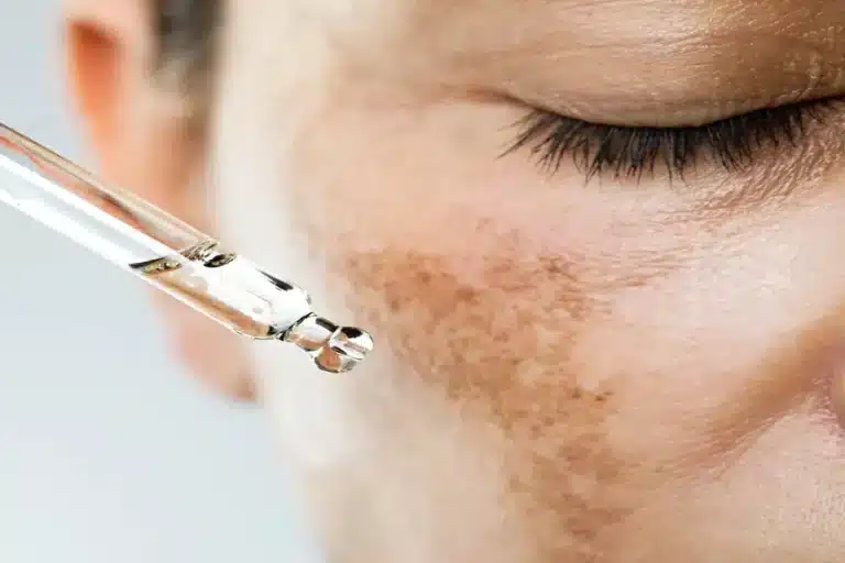
Melasma presents as symmetric brown or gray-brown patches on the face, particularly across the cheeks, forehead, upper lip, and nose bridge. These patches develop gradually over months or years, often triggered by pregnancy, oral contraceptives, or sun exposure. The color remains uniform within each patch, though darkness may fluctuate with sun exposure or hormonal changes.
The patches have defined but feathered edges that blend into surrounding skin. Melasma affects both sides of the face in mirror-image patterns. The surface texture remains smooth without scaling, bleeding, or elevation changes.
Solar Lentigines (Age Spots)
Solar lentigines appear as flat, tan to dark brown spots on sun-exposed areas including the face, hands, shoulders, and upper back. These spots maintain uniform coloration throughout each lesion and sharp, distinct borders. Size ranges from 5mm to 15mm typically, though larger spots may develop after decades of sun exposure.
Age spots remain stable once formed—they don’t grow, change color dramatically, or develop irregular features. The surrounding skin shows other signs of photodamage including fine wrinkles and textural changes. Multiple spots often appear in clusters on areas with highest cumulative sun exposure.
Post-Inflammatory Hyperpigmentation
Post-inflammatory hyperpigmentation (PIH) develops following skin injury or inflammation from acne, eczema, burns, or cosmetic procedures. These dark marks correspond exactly to the previous inflammation site and fade gradually over several months to years depending on depth and skin type. The color ranges from pink to red in lighter skin tones to brown or black in darker skin types.
PIH boundaries match the original injury or inflammation area precisely. The pigmentation remains flat without texture changes or symptoms. Darker skin types experience more pronounced and longer-lasting PIH due to increased melanocyte activity.
Warning Signs Requiring Evaluation
The ABCDE Criteria
Asymmetry indicates concern when one half of a pigmented lesion doesn’t match the other half in shape, color, or thickness. Draw an imaginary line through the center—both halves should appear similar in benign lesions.
Border irregularity presents as edges that appear scalloped, notched, or blurred rather than smooth and even. Melanomas often have borders that fade into surrounding skin or show satellite pigmentation around the main lesion.
Color variation within a single lesion suggests malignancy. Look for combinations of tan, brown, black, red, white, or blue within one spot. Benign lesions typically display one or two shades of brown.
Diameter larger than 6mm (pencil eraser size) warrants examination, though melanomas can be smaller. Any pigmented lesion growing noticeably should be assessed by a healthcare professional.
Evolution represents an important warning sign. Changes in size, shape, color, elevation, or symptoms like itching or bleeding should be evaluated by a healthcare professional. Document lesions with photographs to track changes objectively.
Additional Concerning Features
New pigmented lesions appearing after age 40 deserve careful monitoring, particularly if they differ from existing moles. While new freckles and age spots develop normally with aging, new dark or multi-colored spots should be assessed by a healthcare professional.
The “ugly duckling” sign identifies lesions that look different from other spots on your body. When one stands out as darker, larger, or differently shaped, evaluation by a healthcare professional is recommended.
Symptoms accompanying pigmentation changes increase concern levels. Itching, tenderness, bleeding, or crusting in pigmented lesions suggest possible malignancy. Benign pigmentation remains asymptomatic aside from cosmetic concerns.
Distinguishing Features
Texture and Surface Changes
Benign pigmentation maintains smooth skin texture matching surrounding areas. Melanomas and other skin cancers often develop surface irregularities including scaling, crusting, or ulceration. Run your finger gently across concerning spots—roughness or elevation changes warrant examination.
Seborrheic keratoses, though benign, feel waxy or stuck-on with a crumbly surface. These growths appear suddenly but remain stable once formed. Their appearance includes a well-demarcated border and uniform tan to black coloration despite the raised, rough texture.
Pattern Recognition
Pigmentation from hormones or sun damage follows predictable patterns. Melasma appears symmetrically on both sides of the face. Solar damage concentrates on areas receiving most sun exposure throughout life. PIH corresponds to previous injury sites.
Concerning lesions ignore these patterns, appearing randomly without obvious triggers. They may develop on sun-protected areas or show no relationship to previous skin conditions. A dermatologist can recognize these pattern deviations during examination.
Growth Rate and Behavior
Normal pigmentation develops over months to years, then stabilizes. Melasma may darken with sun exposure or hormonal changes but doesn’t expand beyond typical distribution areas. Age spots accumulate gradually but individual spots remain stable once formed.
Melanomas can grow noticeably within weeks to months. They may develop satellite lesions—small pigmented spots surrounding the main lesion. Rapid growth or spreading pigmentation requires dermatological evaluation.
Clinical Examination Techniques
Dermoscopy
Dermoscopy uses specialized magnification with polarized light to visualize pigmentation patterns invisible to the naked eye. This non-invasive technique reveals structural features distinguishing benign from malignant lesions.
Benign lesions show regular network patterns, uniform globules, or homogeneous pigmentation under dermoscopy. Melanomas display irregular networks, asymmetric globules, blue-white veils, or irregular streaks. These patterns guide biopsy decisions and monitoring strategies.
Wood’s Lamp Examination
Wood’s lamp (black light) examination enhances contrast between normal and abnormal pigmentation. Epidermal pigmentation appears more pronounced under Wood’s lamp, while dermal pigmentation shows minimal change. This differentiation helps determine treatment approaches and expected outcomes.
Melasma classification into epidermal, dermal, or mixed types through Wood’s lamp examination can help predict treatment response. Epidermal melasma may respond to topical treatments, while dermal melasma may require different therapeutic approaches.
When to Seek Professional Help
- Any pigmented lesion changing in size, color, or shape
- New dark spots appearing after age 40
- Lesions with multiple colors or irregular borders
- Spots that bleed, itch, or become tender
- Pigmentation that looks different from your other spots
- Asymmetric lesions or those larger than 6mm
- Spots developing on palms, soles, or under nails
- Pigmentation not improving after 3 months of treatment
- Family history of melanoma with new pigmentation changes
- Previous skin cancer with new concerning lesions
Commonly Asked Questions
How quickly should pigmentation changes be evaluated?
Rapidly changing lesions require evaluation within days to weeks. New spots in adults over 40 or any lesion meeting ABCDE criteria deserves assessment within one month. Stable pigmentation without concerning features can be monitored at routine skin checks.
Can smartphone apps accurately diagnose skin lesions?
While apps may help track changes over time, they cannot replace professional evaluation. Accuracy varies widely between applications, and many concerning lesions are misclassified. Use apps for documentation but rely on dermatological examination for diagnosis.
What happens during a pigmentation assessment?
Dermatologists examine lesions visually and with dermoscopy, reviewing the entire skin surface for other concerning spots. Suspicious lesions may require biopsy for definitive diagnosis. Photography documents lesions for future comparison. The consultation includes discussion of findings and treatment recommendations.
Can normal pigmentation transform into cancer?
Most benign pigmentation remains benign permanently. However, any pigmented lesion can theoretically undergo malignant transformation. Regular monitoring identifies concerning changes early when treatment outcomes may be favorable.
Next Steps
Distinguishing benign pigmentation from serious conditions requires careful observation of ABCDE criteria and prompt evaluation of suspicious lesions. Document concerning spots with photographs and monitor for rapid changes.
If you’re experiencing asymmetric pigmentation, lesions with irregular borders or multiple colors, or spots that are changing rapidly, an MOH-accredited dermatologist can provide comprehensive skin examination using dermoscopy and other diagnostic techniques.


